


Scan It! Magnifying the Potential of Our Collections
How digital imagery is advancing research and creating global scientific collaborations

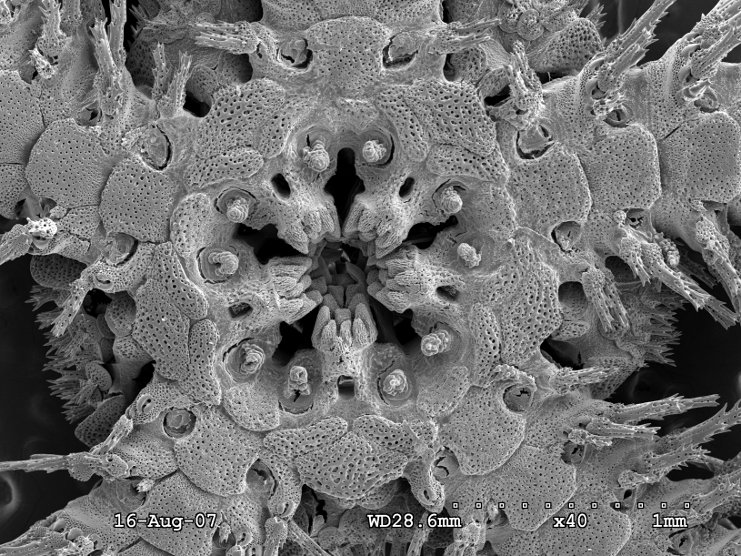
This is a SEM image of the brittle star Amphiodia urtica. Gordon Hendler, Curator of Echinoderms explains “there are 30 large, radially arranged skeletal plates covering the central “disk” of this baby brittle star’s body. As the animal grows, new plates arise in an orderly symmetrical pattern, and they number in the thousands in an adult.” This scan allows us to see these skeletal plates in great detail.
By Bridget Altman and Tyler Hayden
Under the Microscope
Try to imagine the smallest animal you’ve ever seen with the naked eye. Got it? Okay, what does its mouth look like? Minute anatomical details like mouth shape can reveal exactly how the smallest creatures around are staying alive, and lets us get a better view of little things that can help us see the big picture.
This SEM image captures the central disk and five-jawed mouth of the brittle star Ophiothrix spiculata, one of 7,000 images Dr. Hendler used in a three-year study of anatomy, function and development of brittle star mouthparts. His research, which culminated in a 123-page monograph, hinged on SEM. Images like this helped Hendler and his colleagues learn that there are two different types of brittle star mouths: one for species like predators and another like the funnel-shaped one here, used to compact and crush microscopic particles of food. The pictured mouth is about 2mm across, or roughly half the size of Lincoln's head on a penny.
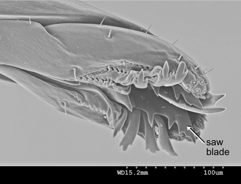
When NHM’s Entomology Curator Brian Brown and Collections Manager Giar-Ann Kung were doing field work in Brazil, they noticed a phorid fly in the Dohrniphora longirostrata-group was leaving some ants headless. Kung suggested the ants were being decapitated, but Brown was initially skeptical. A closer look at the fly’s proboscis (mouthpart) under NHM’s Scanning Electron Microscope showed the structure of the mouthparts that helped support Kung’s observation. Getting a closer look at the proboscis revealed nature’s tiny saw blade.
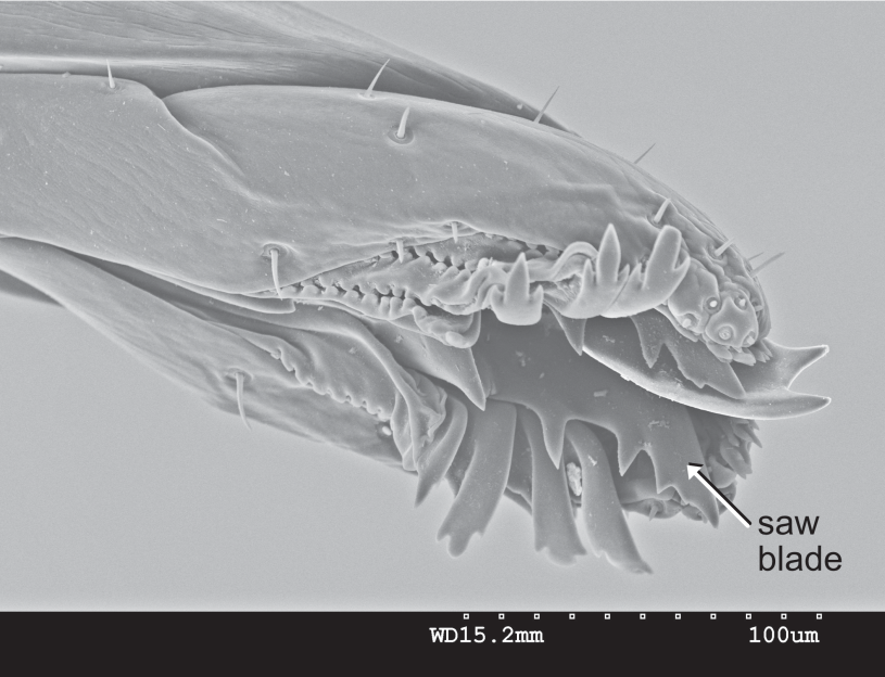
Image by Giar-Ann Kung, Entomology Department
The (above) SEM depicts a Dohrniphora longirostrata-group proboscis, mouthpart, (about 900 microns (μm) per the scale bar bottom right) collected Brown and Kung in Brazil, and (below) magnified to about 200 μm. For perspective, a strand of human hair is about 70 μm, plus or minus 20 μm depending on thickness. This fly uses its proboscis with that serrated appendage to saw off the heads of its prey.
“When we can see more of the super small things, we get a better
picture of the specimen; we get a new story to share.”
—Dr. Trina Roberts, Associate Vice President for Collections
PUTTING THE ELECTRON IN S-E-M
Scanning Electron Microscopes have a more powerful magnification ability than other microscopes thanks to their use of electrons. While traditional microscopes use photons (particles of light) the SEM uses electrons, negatively charged particles that have much shorter wavelengths than photons. These shorter wavelengths help reveal much finer details. It’s like the first time you watched a sporting event televised in HD and suddenly you can see the blades of grass on the field.
This type of imaging has the power to help researchers understand their subjects on a deeper level, and the images they produce can help inspire wonder in our natural world. So when NHMLAC received a grant from the National Science Foundation to help fund the purchase of a new SEM, NHMLAC’s Research and Collections team was thrilled. “The new SEM is an indispensable modern research instrument that will allow our researchers to view specimens and artifacts at tremendous magnifications and with superb resolution,” explains Dr. Jody Martin, Associate Vice President for Research. “Equally important, the SEM will be displayed for students and visitors to NHM to allow them to see "science in action" and learn about the wonders of the microscopic world as they watch our scientists at work.”
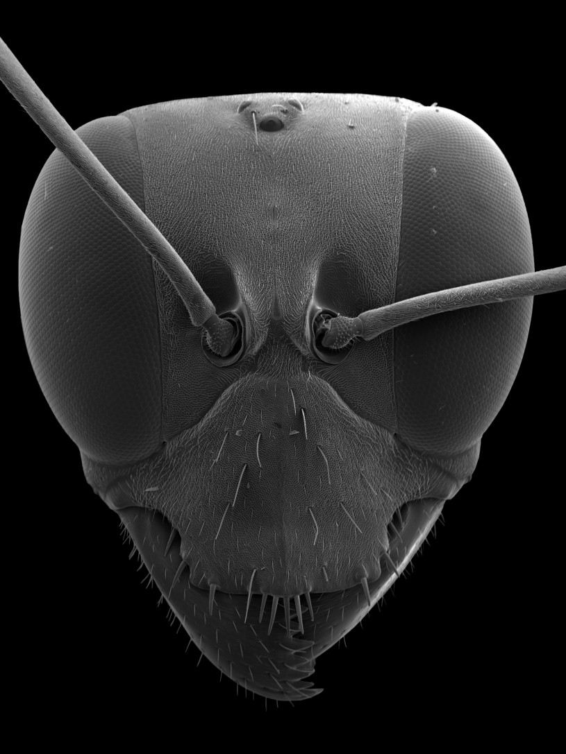
Gigantops destructor are big (for ants), at around 7mm long (their heads are around 2mm wide) but this SEM image reveals the powerful jaws and distinct eye in powerful detail.
With even more detailed images employing many different techniques, NHMLAC researchers can bring their science to a smaller magnitude, revealing whole new dimensions of understanding and wonder. The increase in digitization, creating digital catalogues of data from specimens, has allowed our researchers to examine different facets of tiny specimens. Dr. Trina Roberts, Associate Vice President of Collections, notes that “when we can see more of the super small things, we get a better picture of the specimen; we get a new story to share.”
CT Scanning
For nearly 50 years, Computed Tomography scans, more commonly known as CT scans, have been used to help doctors get a look at our brains without cracking our skulls. Now, this same type of technology lets museum scientists get a better look at priceless specimens without damaging them, and it has the added bonus of making those one-of-a-kind collection items available to scientists around the world.
LITTLE FISH ARE KIND OF A BIG DEAL
Thanks to a National Science Foundation grant, Dr. Bill Ludt, NHMLAC’s Assistant Curator of Ichthyology, is hoping to get a good look at cryptobenthic fishes. Just so we’re on the same page: “crypto” refers to “cryptic” or “mysterious” and “benthic” meaning “living on the seafloor”. Ludt explains, “Even though it is not part of their name, one of the defining characteristics of cryptobenthic fishes is their small size.” Using CT scanning, Ludt is able to get a new look at these tiny reef-dwelling swimmers.
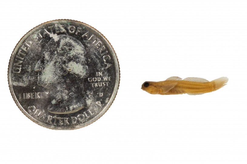
CT scanning helps researchers overcome the diminutive size of cryptobenthic fishes like this Risor ruber
“There are 17 different families of cryptobenthic fishes, around 6,000 species, and they make up 10 percent of all vertebrate diversity,” Ludt says. Not fish diversity, one tenth of the diversity of all vertebrates, everything with a backbone. What’s more, they play a crucial role in the productivity and biodiversity of reefs. While phytoplankton is the well-known base of the oceanic food web off the coastal waters of places like Southern California, cryptobenthic fishes play a crucial role in the food webs of reefs, and scientists are only beginning to understand the key role these fishes play in supporting the spectacular diversity and productivity of coral reefs.
Cryptobenthic fishes have a short lifespan and produce a lot of larvae. It’s a live-fast, die-young lifestyle. Ludt stresses that while they don’t replace phytoplankton, larvae and adults of these cryptic critters are responsible for approximately 40 to 50 percent of food chain activity in reefs, but “because these fishes are so small, looking at their internal anatomy is extremely difficult, making it hard to understand how they fit into their environment and how they’ve evolved”.
There is a very high level of expertise needed just to examine and dissect the specimens, and often the fragile specimen gets destroyed in the process. High resolution imagery, like CT scans, will help researchers all over the world see these creatures with new eyes. “We’re going to scan their skeletons as well as their soft tissues, such as organs, veins, muscles, and post the scans publicly for everyone to use,” encouraging global collaboration.
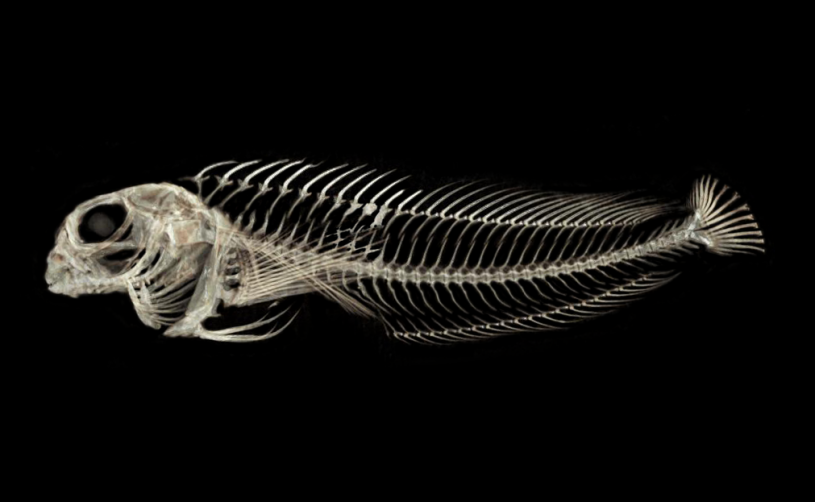
This CT scan of a Rockpool Blenny (Hypsoblennius gilberti), a native of Southern California, has had its soft tissues digitally removed, illustrating the exquisite precision of its minuscule skeleton.
This is particularly beneficial now since the pandemic has limited in-person access to museum collections, Ludt points out: “Researchers can’t visit our collections right now, but they can still get research done by downloading data. Scanning and publishing data on these specimens increases the equity and reach of our science.”
3D Scanning
Another important way NHMLAC scientists are making collections more digitally accessible is through uploading 3D scans of specimens online. These high resolution images, paired with the dynamic angles of a 3D scan, are great tools of scientific investigation.
A NEW KIND OF RECORD AT LA BREA TAR PITS
Emily Lindsey, Associate Curator and Excavation Site Director at La Brea Tar Pits along with LBTP staff and colleagues from UCI, Vanderbilt, Loyola Marymount, Marshall and Des Moines University are working on a project that involves sampling hundreds of jaw bones from dire wolves, saber-toothed cats, coyotes, horses, and bison, to understand how these species were impacted by, and adapted to, Ice Age climate changes. To determine the exact age of the fossil bones, the team takes a small sample of bone, treats it with chemicals and then burns it—a process scientists call “destructive analysis.” Before that destruction begins, they use a 3D surface scanner to create exact digital replicas. The blue light scanner is connected to a high-powered laptop loaded with the scanning software. The fossil is placed on a turntable and the scanner is aimed at each side of the fossil to record all angles. Each scan is then aligned and merged together to create a 3D mesh, which is then fused into a solid model with texture and color.
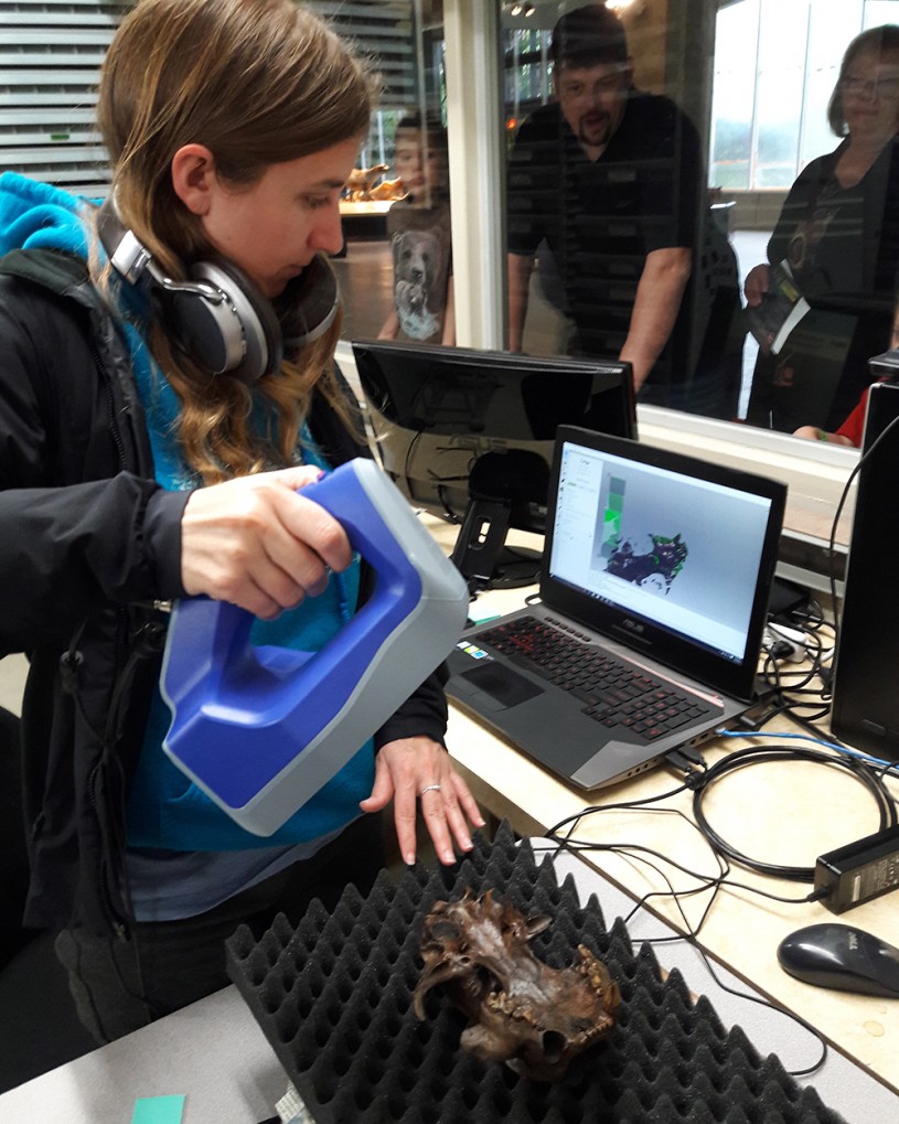
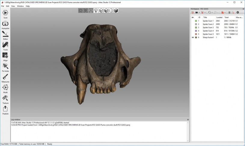
Fossil Preparator Carrie Howard is taking a 3D scan of a fossilized Mountain Lion, Puma concolor in front of visitors at La Brea Tar Pits. The fossil is safely placed on a turntable and the scanner is plugged into a computer with the scanner’s software. The scanner is aimed at a proper distance and flashes light at the fossil. The cameras and software record the 3D data.
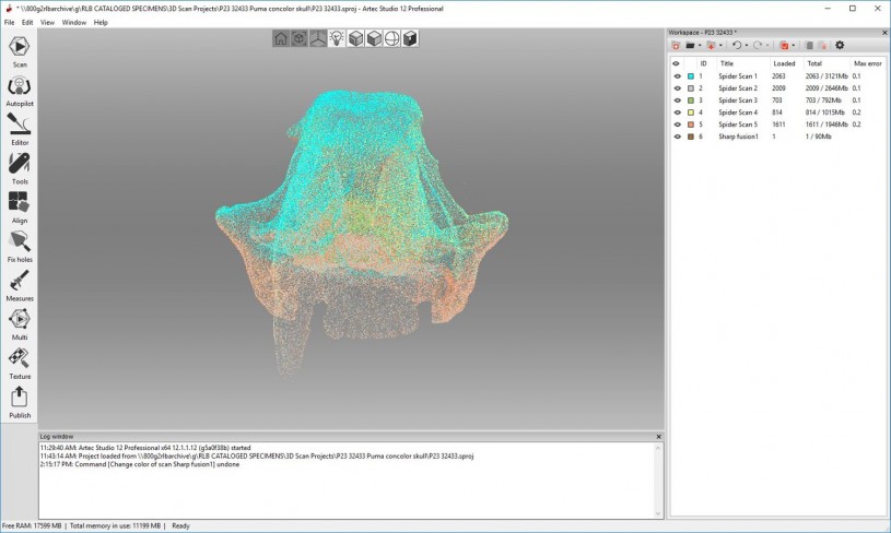
Multiple scans are merged together in the software to create a 3D mesh model of the fossil.
After the solid model is processed from the mesh, and the texture and color are captured from the scanner and rendered over the solid model, you get a finished 3D scan replica of the fossilized Mountain Lion Puma concolor.
Aisling Farrell, Collections Manager at La Brea Tar Pits, explains that most of the collections are not easily accessible to people all over the world, so these scans open up a new path for researchers and students to access data from our records of specimens. For example, Farrell recalls a group of students from Loyola Marymount University were working on a project before the pandemic began that involved taking measurements from fossil specimens at La Brea Tar Pits. Thanks to the 3D scans, these students were able to continue their research safely from home. Farrell exclaimed “this technology truly magnifies the potential of our collections.”
Let's Get Digital
Scroll down within the interactive window below to zoom in, scroll up to zoom out, and be sure to check out the rest of La Brea Tar Pits' 3D digital collection.
Explore even more 3D models from La Brea Tar Pits and NHM's Invertebrate and Vertebrate Paleontology collections on our SketchFab account.
A Home For The Data
Now, let’s talk data. Once we have all of these amazing scans and images, what happens to them? Many of NHMLAC’s Curators and Collection Managers have been sharing digital collections data to the Global Biodiversity Information Facility, or “GBIF” . GBIF is one of several online portals that aggregate and combine biodiversity data from research institutions around the world for museum scientists, students, educators, and the public, giving NHMLAC’s specimens significantly increased visibility. Researchers from around the world can browse records of NHMLAC specimens using aggregators like GBIF to identify specimens in their own collections or even discover a new species (browse NHMLAC's digitized collections in GBIF, its American equivalent iDigBio and more here).
With support from the National Science Foundation, Invertebrate Paleontology staff worked collaboratively from 2017 to 2020 with a team from La Brea Tar Pits to digitize several large collections of fossil insects. Some of these specimens have been in the collections for nearly a century, and have remained in drawers, untouched since their excavation. There are millions of specimens in the Invertebrate Paleontology and Rancho La Brea collections, and it is virtually impossible to image each individual fossil, so this process is highly selective.
Specimens as small as insects are especially time-consuming to image. This was accomplished by the Invertebrate Paleontology collections using a macrophotography technique called “focus stacking” whereby collections staff and volunteers must take a series of images of for each tiny fossil selected for imaging. Later, the images are stitched together using a computer software program to create one high-resolution winner. Uploading even a few well-preserved and important specimens is worth the effort.
Austin Hendy, Curator of Invertebrate Paleontology, explained “two decades ago when museums first started to share images of their specimens online, there was concern among collections staff that people wouldn’t visit physical collections anymore. But the opposite has been true! It turns out, if you put your data out there, people can see what you have, and they will come”. But making specimens available online leads to new collaborations too.
In fact, a researcher from Italy was browsing GBIF and noticed several unusual beetle specimens that were digitized from NHMLAC’s collection of fossil insects from Germany. After reaching out to the Collections Manager, Lindsay Walker, about these specific specimens, Walker sent the researcher more detailed images to review. Using the images from GBIF, as well as additional imagery provided by Invertebrate Paleontology, the researcher described several new species of beetles, including three named for volunteers in the Invertebrate Paleontology collections.

A digitized fossil specimen of Cantharis lidiae, a beetle named after Lidia Lustig, an avid geologist and long-time NHMLAC volunteer.

A digitized fossil specimen of Cantharis bradburyi, a beetle named after David Bradbury, a generous and active NHMLAC volunteer, life-long enthusiast of fossil arthropods, and active member of the Southern California Paleontological Society.

A digitized fossil specimen of Rhagonycha carolynae, a beetle named after Carolyn Weiss, a dedicated volunteer at NHMLAC with a passion for museum education, research and collections.
Advancements in scanning technologies and their greater use are expanding access to collections, but these emerging technologies also offer their own challenges. Enormous files and the need for specialized software and equipment to view and interact with them mean not all images and scans can be shared or stored easily in the same places. For museums, it is crucial to maintain links between collection information stored in different ways and on different platforms, meaning innovations in managing the flow of information have also been needed. . While new and improving scanning technologies offer new challenges, their promise remains great. Roberts explains “now more than ever, various types of imagery can help augment the collections through research potential, accessibility and outreach, encouraging collaboration and advancing science, which magnifies both the importance and reach of every specimen.”


