Small Wonders
NHM’s new Scanning Electron Microscope Lab connects guests with big science and tiny specimens.

Deep in the Gem and Mineral Hall, there's a portal to a place of microscopic wonders—and it glows blue. NHM's SEM Lab lets visitors share in the hidden worlds all around us as they watch researchers investigate.
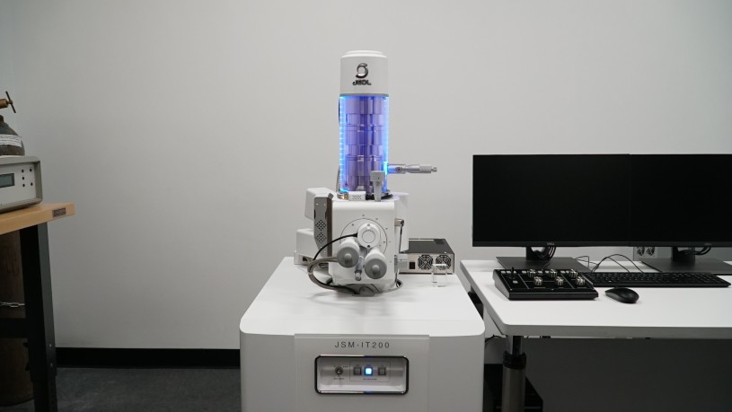
At NHMLAC, scientists explore our natural and cultural worlds in awesome ways every day, but most of that work happens behind the scenes. The new Scanning Electron Microscope Lab (or SEM Lab for short) in the Gem and Mineral Hall brings some of that mind-blowing exploration to Museum visitors, opening doors to incredible tiny new worlds with big scientific impacts.
Whether it’s figuring out the raw material of an ancient tool or the invisible makeup of crystals, knowing the chemical makeup of a plant, animal, or mineral can contribute to our scientific understanding of the objects we study. Combined with its significant power of magnification and increased resolution of microscopic objects, the SEM is an invaluable tool for research across the Museum.
SEMs work on a whole different level from the light microscopes you might be more familiar with, achieving mind-blowing magnification with subatomic particles. “How someone ever came up with that is absolutely astounding to me because it really is almost like black magic,” says Kirk Fitzhugh, NHM’s Curator of Polychaetes and the Museum’s resident SEM sage. Dr. Fitzhugh was a superuser of NHM’s old SEM—an impressive and invaluable research tool that was inaccessible to Museum visitors. “Anytime I would bring people down to see it in operation, they were just invariably fascinated. And then many of them would ask, ‘Why isn't this visible to the public?"
When the old SEM became unusable, Fitzhugh and other NHM researchers saw an opportunity. Having such an instrument available for scientific research be more visible could make it a powerful tool for inspiring wonder among Museum visitors, and that same visibility could help attract funding from philanthropic individuals and organizations. Along with being incredibly high-powered, SEMs are incredibly expensive. With a successful grant from the National Science Foundation and funding from the Ludwick Family, the SEM Lab has been inviting visitors into miraculous microscopic realms since September 2022.
Seeing Without Light
While visitors can watch NHM researchers working on specimens and see the amazing images they produce, SEMs actually work without using light or anything in the visible spectrum at all.
When a sighted person “sees” an object, light is hitting that object—some of the light is absorbed, but some is reflected, and when that reflected light hits our eyeballs, they alert our brain, and ta-da! We "see" that something. Light microscopes basically do this with the addition of glass lenses to magnify the objects we want to see better. These microscopes either pass light through the specimen or reflect light off it to help us get a better look. The highest magnification you can achieve with a good light microscope is about 1,200 times–but some SEMs can magnify things more than 100,000 times. And they do it in the dark.
Instead of visible light, scanning electron microscopes use, well, electrons—negatively charged particles. They’re complicated! But what we need to know is that their wavelengths are much, much smaller than the visible light photons (the light you’ve just been reading so much about) used with light microscopes. An SEM produces a focused beam of electrons that is directed at specimens, causing other “secondary” electrons from surfaces of the specimen to be emitted. Those electrons are then collected by a sensor that converts them into the images we see. It is the vastly smaller wavelengths of electrons that enable us to achieve such high magnifications and observe greater details compared to using visible light.
Small Wonders, Big Science
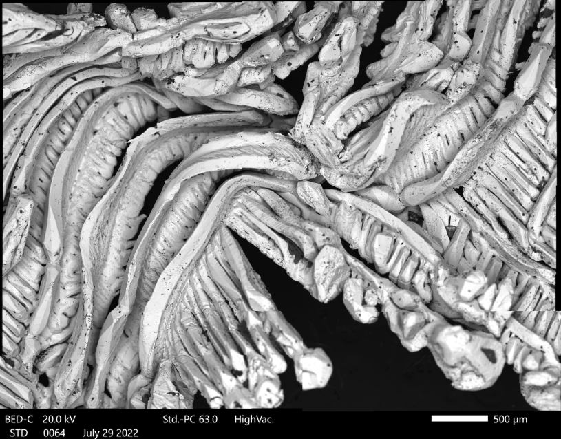
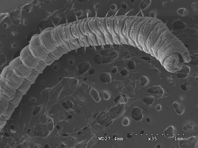
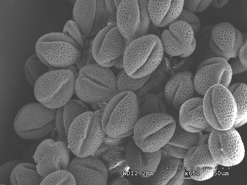
SEM images are strikingly tactile, finely-detailed topographical maps of whatever’s viewed with the scope. These images are more than just amazing pictures: ant heads reveal all their intimidating bumps and powerful jaws, the serrated edge of a fly’s mouth becomes clear (explaining how it can saw off ant heads), and the structure of feathers helps researchers understand more than ever about how birds achieve their striking colors.

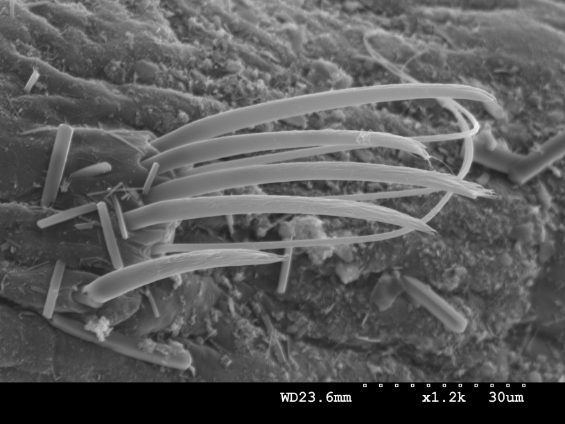
“Right now we're using the SEM to basically get the 3D structures of feathers’ microstructures, an aspect that's been largely ignored for a really long time. We can actually zoom in further and get at how the molecules within the feathers are arranged as well,” says Allison Shultz, Curator of Ornithology and feather fanatic. Pictured above, an SEM image of a Tangara chilensis' rump dorsal feathers.

SEM can help us to describe the entire feather versus just what we can see with our human eye or even with the confocal microscope (a powerful light microscope that uses lasers to measure fluorescence),” says Shultz. A Tangara chilensis feather with no magnifcation.

iNaturalist Photo 158016451, (c) Abby Darrah, some rights reserved (CC BY), uploaded by Abby Darrah tangara
A Tangara chilensis or paradise tanager in the wild. The how and why of these colorful bird feathers is a secret locked in their microstructure, which is only visible in using SEM.
“With a light microscope, visible light is passing through or reflected off a specimen. That light offers a limited range of magnification and resolution. With an SEM, all you're seeing are surface details of objects because the electrons are only hitting the surface and then image formation is produced due to the interactions of those electrons with the surface atoms or molecules,” Kirk Fitzhugh
Those highly detailed images of very small things are especially valuable, but they’re just one way SEM can tell us more about specimens–it can also reveal their chemical building blocks. The beam of electrons bombarding a specimen is strong enough to knock electrons out of their designated locations in atoms. You know what? We’ll let Dr. Fitzhugh explain:
“If an electron gets thrown out, an electron has to replace it. So another electron within the atom moves into that location. When that happens, it releases energy in the form of X-rays. And the type of X-ray is almost like a fingerprint that tells us what elements are in the specimen. Is it an atom of gold, an atom of copper, or an atom of calcium? The x-rays have a distinctive signature that tells you what element—or set of elements—makes up the object you're examining.”
This type of analysis can inform researchers about the compositions of structures of the tiniest organisms or inanimate objects such as minerals.
Fantastic Voyages Into Everyday Things
SEM has the power to open up unbelievable views of fascinating research and also exposes wonder in the world all around us that we simply can’t see without it. Recently Dr. Fitzhugh introduced some lucky visitors to the fantastic side of a common ingredient in a ton of household goods. Diatomaceous earth is in everything from aquarium filters to bath mats and insecticide powders. To our eyes, it just looks like white powder, but with SEM we can finally see what we’re really looking at. “So this powder is actually billions of skeletons of single-celled marine algae called diatoms. They produce a skeleton made of silica and when they die, the skeletons sink to the bottom [of the ocean] and they form huge layers of rock.”
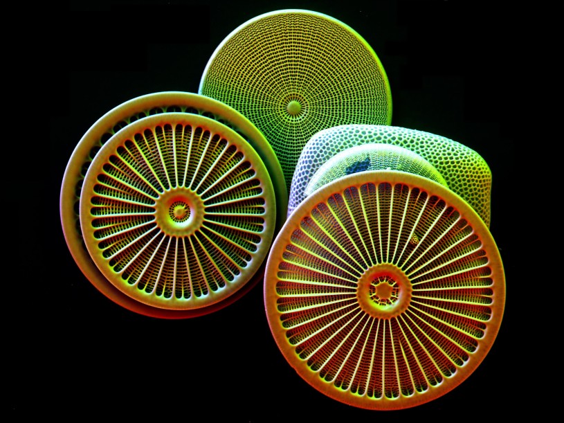
Under the SEM’s magnification, this ordinary household item reveals its extraordinary beauty. “You're going from just this incredibly fine powder, go up to a magnification of about 5,000 times, and you're seeing these intricate structures,” says Dr. Fitzhugh. The artificially colored skeletons of diatoms above showcase SEM’s ability to bring the everyday wonders of our world we could never see—and the crucial roles they play on our planet beyond our sight. “Ecologically they're very, very significant,” says Dr. Fitzhugh. “About 20 percent of the oxygen produced on this planet is produced by diatoms.”
Helping visitors understand the big impact seeing really tiny things can have on science is only getting started. Dr. Fitzhugh and other NHM researchers plan to showcase the stupendously small side of more common objects in the future. Right now, school groups can watch Museum researchers working on the live feed on a large screen monitor. In the future, the SEM Lab will have an intercom system so researchers can talk guests through whatever microscopic world they’re exploring—and guests can ask questions as they journey through the wondrous wee world of SEM.
